
ミイラは2000年以上前のもので、英国スウォンジー大学のエジプトセンターのコレクションとして保有されていた。

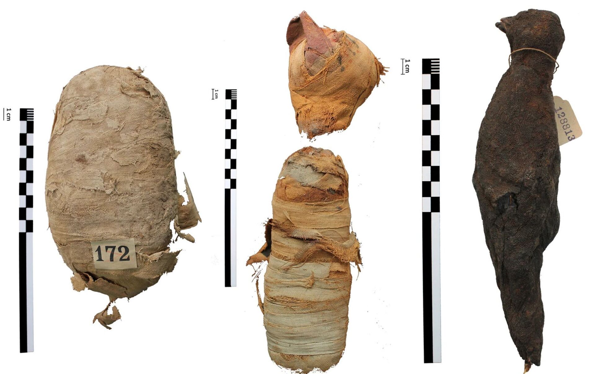
蛇のミイラ

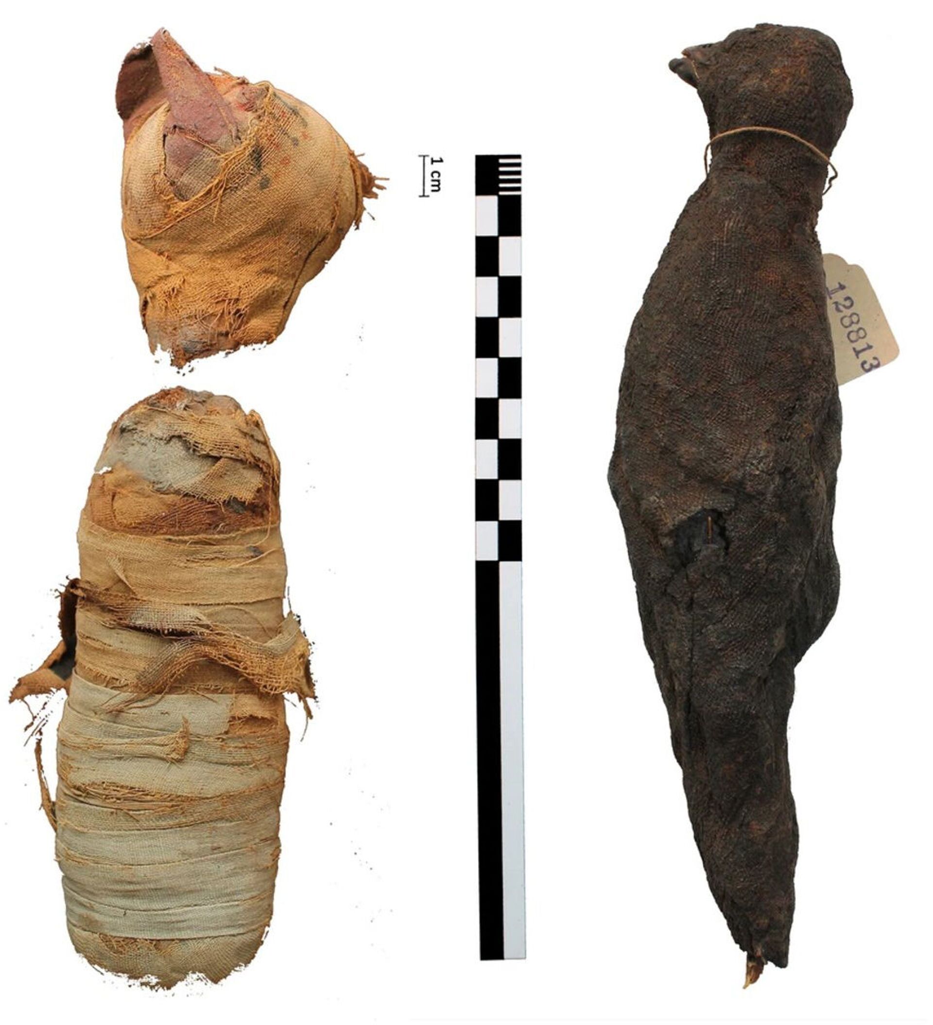
鳥のミイラ

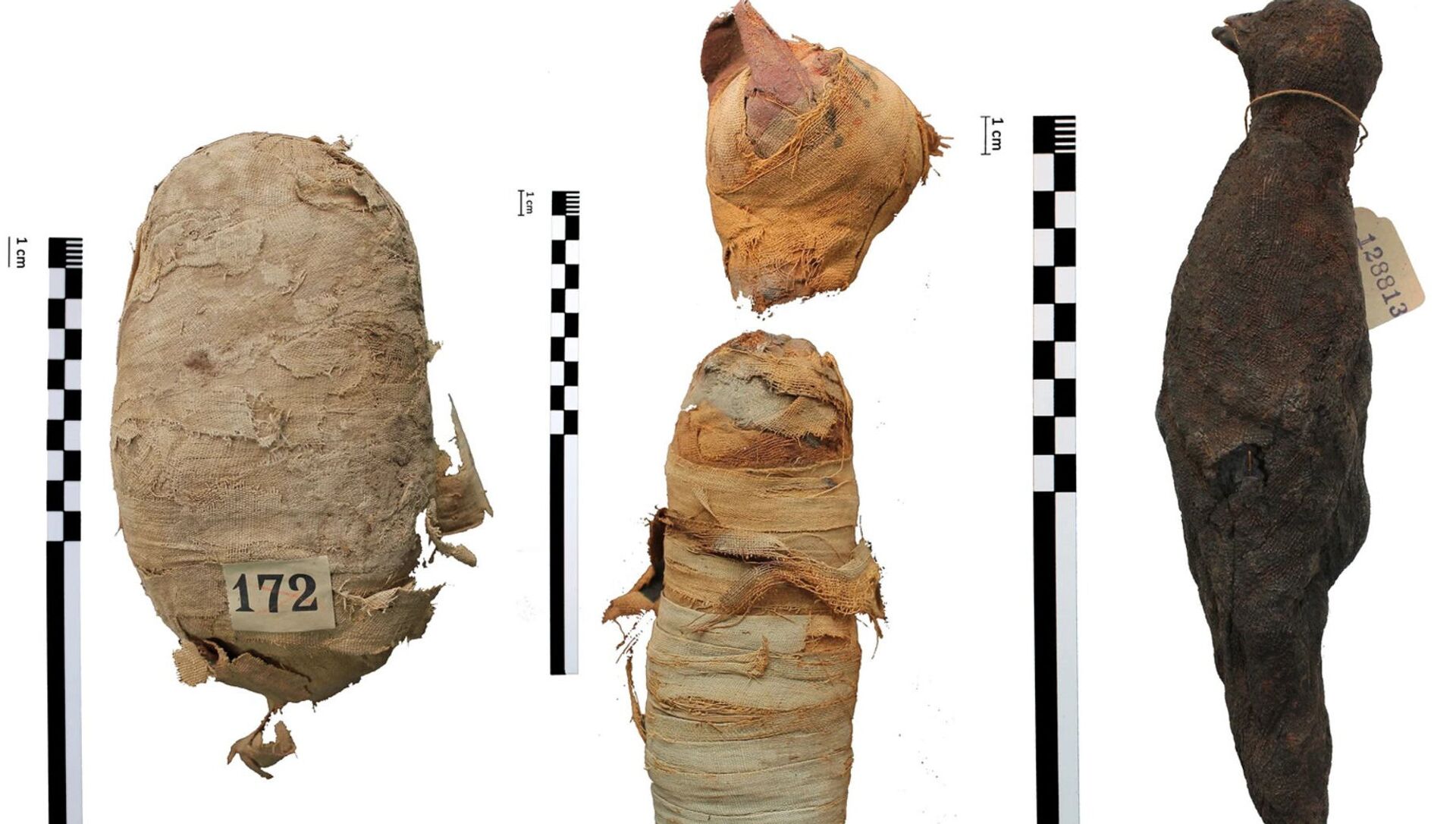
猫のミイラ

猫のミイラ
蛇のミイラ
鳥のミイラ
猫のミイラ
猫のミイラ

3D画像での撮影によってミイラの解析度は高く、研究者らは、ミイラの1体が5ヵ月未満の子猫であることを確認した。子猫はおそらく窒息死している、鳥は猛禽類に属し、また、蛇は若いエジプトのアスプコブラだと判明した。蛇の腎臓の損傷跡は、生存期間を通じて脱水症状であったことを示している。蛇は神への生贄として供えられ、骨が砕かれていることから殴打によって死亡している。
古代エジプト人は、猫や蛇、ワニ、犬などをはじめ、動物をミイラ化していた。時には飼い主とともに埋葬されたが、しかし、多くの場合、貢物として捧げられ、その後、司祭が遺骸を防腐しミイラとした。こうして約7000体の動物のミイラが作られたと考えられている。


